Re: One in Six Americans Have Had H1N1; 10,000 Have Died
These are the actual notes from a pathologist emailed to me and printed with permission. Edited for readability. Bolding mine.
Please take heed. There is a very dangerous side to Swine Flu.
Florida1
(Sharon Sanders)
"We did an autopsy last week on a fellow who died of swine flu. We got the laboratory tests back from our swabs, but he seemed to fit? I will just read ... out the circumstances of the death because I think it's a pretty typical story.
The guy was aged 47 years.
He died on July 14. but six days before on July 8, he had started to feel unwell with flu-like symptoms of sore throat, joint pains and headaches. Then he was coughing and had a loss appetite.
Those symptoms continued until the Sunday, July 12. He then started to cough up a blood. He had a persistent cough and on the Monday night he was observed by his family members to have difficulty breathing. Then the next day, around 10 o'clock, he was found dead in bed. An ambulance was called, but he was already dead. A fairly short but typical length of illness of about 6-7 days.
He was not diabetic but as far as I'm aware just hypertensive (and over weight).
I will just go to the heart of it. And that's to do with the lungs. There was a little bit of straw-colored fluid around the lungs in the chest cavity, which is a non-specific finding, but when the doctor who I was supervising doing to case was looking at the airways, the trachea and the bronchi, and they were really red, like tomato, beetroot red. They are normally a light-pink color, so clearly inflamed. And this was all the way down the bronchiole tree.
People who die of bronchi pneumonia or standard pneumonia, you will often see pus in the airways, but there was no pus in this guy's airways, just intensely red.
The histology of the airways showed a lot of epithelial ulceration. A loss of the surface lining cells with obviously bleeding underneath, which I think partly explains why he was coughing up blood. And acute inflammation of the upper air passages -- the trachea and the main bronchi. But not of the sort that looks like acute bronchitis with neutrophils. Neutrophils are what's colloquially called pus cells. And if you have got acute bronchitis from say a bacteria like staph. or something like that, they are the first line of defense cells that go and gobble up the bacteria.
In this particular case, there were hardly any neutrophils because it's not a bacterial infection, it's a viral infection.
Not much evidence of a secondary bacterial infection at all.
Which is a little surprising, but that?s what we saw. There were hardly any neutrophils. There were some floating around, but the main cells were lymphocytes which are more to do with chronic inflammation and viral infections as well. And other forms of acute inflammatory cells as well called eosinophils and they named that because of the intense orange color of the cytoplasm of the cells when you stain them with a particular stain.
But their function is more related to allergy. So if you see a lot of eosinophils in lungs in particular, you might think of asthma or some allergic phenomenon that's attracting those eosinophils to that site. I am not suggesting for a minute that this was an allergic phenomenon at all. This was a viral-type process. For whatever reason, and I don?t understand it, those sorts of cells -- the eosinophils and the lymphocytes were being attracted to that inflammatory area.
That was the first thing -- so intense inflammation of the upper air passages. The lungs were heavy. They were about twice to three times as heavy (as normal). The left lung is normally less than the right because it has got two lobes and the right lung has got three lobes. And the left lung in this case weighed 630 grams and the right lung weighed 830 grams. The left lung is generally about 200-250 and the right about 350-400 or so. At least double normal for each.
All the organs in large people tend to be bigger anyway, particularly the liver and heart. The lungs not so much. Those weights are pathological of something, they are not standard weights.
There is supportive tissue in the lungs, but the bulk of lungs is air. And there shouldn?t be any bleeding into the tissue of the lungs, but what we were seeing was precisely that. We were seeing areas of what looked to be hemorrhage throughout both lungs, particularly in the lower lobes.
That again is quite different to your standard bacterial pneumonia, where you see pus in the small airways and you see what's called consolidation of the lungs by lobar pneumonia say from an ordinary bacterial organism that you might acquire at home or in the community. We saw those abnormalities in conjunction with the upper airway inflammation.
We knew we were onto something which was not standard pneumonia and the story of course was of a flu-like illness and he fitted the bill with him being at risk for swine flu.
So we were hot on the trail of that and we took swabs of the trachea, of the lung and the nasopharynx and I think they all came back positive for swine flu.
That in short are the findings.
Again, consistent with viral pneumonia where there are not many neutrophils but there are a lot of lymphocytes, mainly lymphocytes in the alveoli spaces, those grapelike sacks that do the gas exchange. A lot of chronic inflammatory cells, mainly lymphocytes, some eosinophils and little aggregates of neutrophils.
If you were to show this to a pathologist blind without any story, this should not be regarded as a standard pneumonia. It really shows the typical features of what would be a viral pneumonia; some sort of viral lower respiratory tract infection.
And then we looked at the heart. Sometimes with a viral infection you can get what's known as myocarditis, which is inflammation of the heart basically. There was no evidence that this fellow had that.
...
(We) couldn?t see any inflammation anywhere else except maybe the spleen. That?s not surprising because it has a lymphoid immunological function anyway. But in a viral infection, you get very non-specific changes in the spleen.
This really was upper and lower air passages and nowhere else.
The viral pneumonia, the virus (is) basically nuking the alveoli and causing severe hypoxia.
Yes, I think it?s that. This guy never made it to ICU so I don't have any of those parameters. I am only guessing, but I think it was partly that and also the effects of a viremia -- just having a pretty virulent virus spreading rapidly through the body and particularly being attracted to a very important site like your lungs makes people profoundly unwell. And on a background of these people having diabetes, high blood pressure, obesity and so on.
Viral pneumonia, I said. I don?t know what the mechanism is. Probably hypoxia eventually because of the inflammation in the lungs -- there was a lot of that.
(Pulmonary hemorrhage) was part of the inflammatory process.
We're on a learning curve here. we don?t know what the pathology of swine flu actually is when it gets down to it. If you recover from it, fine. Nobody is going to biopsy your lungs after you have made a recovery. It's only when people die and they are submitted to autopsy that people like me take over the case and start looking at their lungs.
The sorts of people we really won't be autopsying because their cause of death is known, are the people who are on ventilators in ICU. We are seeing a different population -- people who die at home of unknown causes or where the GP won't sign a death certificate. They don't come to the ICU's attention.''



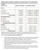


 . I'm just saying that for the next four weeks we're going to hear from every news report and newspaper that H1N1 has killed 10,000 in the US, but on the day the estimate is issued, it's already under by 6000 fatalities. Ah well.
