Re: Nature - Human host factors required for influenza virus replicationnear-final version
Re: Nature - Human host factors required for influenza virus replicationnear-final version
ATPase
From Wikipedia, the free encyclopedia
Jump to:
navigation,
search
<!-- start content -->
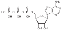
 Adenosine triphosphate
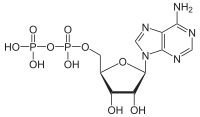 Adenosine diphosphate
ATPases
Adenosine diphosphate
ATPases are a class of
enzymes that
catalyze the
decomposition of
adenosine triphosphate (ATP) into
adenosine diphosphate (ADP) and a free
phosphate ion. This
dephosphorylation reaction releases
energy, which the enzyme (in most cases) harnesses to drive other
chemical reactions that would not otherwise occur. This process is widely used in all known forms of
life.
Some such enzymes are
integral membrane proteins (anchored within
biological membranes), and move
solutes across the membrane, typically against their concentration gradient. These are called
transmembrane ATPases.
<table id="toc" class="toc"> <tbody><tr> <td>
Contents
[hide]
</td> </tr> </tbody></table> <script type="text/javascript"> //<![CDATA[ if (window.showTocToggle) { var tocShowText = "show"; var tocHideText = "hide"; showTocToggle(); } //]]> </script>
[edit] Functions
 Na<sup>+</sup>/K<sup>+</sup>ATPase
Transmembrane ATPases import many of the metabolites necessary for
cell metabolism and export toxins, wastes, and solutes that can hinder cellular processes. An important example is the sodium-potassium exchanger (or
Na<sup>+</sup>/K<sup>+</sup>ATPase), which establishes the ionic concentration balance that maintains the
cell potential. Another example is the
hydrogen potassium ATPase (H<sup>+</sup>/K<sup>+</sup>ATPase or gastric proton pump) that acidifies the contents of the stomach.
Besides exchangers, other categories of transmembrane ATPase include
co-transporters and pumps (however, some exchangers are also pumps). Some of these, like the Na<sup>+</sup>/K<sup>+</sup>ATPase, cause a net flow of charge, but others do not. These are called "electrogenic" and "nonelectrogenic" transporters, respectively.
[edit] Mechanism
The coupling between ATP hydrolysis and transport is more or less a strict chemical reaction, in which a fixed number of solute molecules are transported for each ATP molecule that is hydrolyzed; for example, 3 Na<sup>+</sup> ions out of the cell and 2 K<sup>+</sup> ions inward per ATP hydrolyzed, for the Na<sup>+</sup>/K<sup>+</sup> exchanger.
Transmembrane ATPases harness the chemical potential energy of ATP, because they perform
mechanical work: they transport solutes in a direction opposite to their
thermodynamically preferred direction of movement?that is, from the side of the membrane where they are in low concentration to the side where they are in high concentration. This process is considered
active transport.
For example, the blocking of the vesicular H+-ATPAses would increase the pH inside vesicles and decrease the pH of the cytoplasm.
[edit] Transmembrane ATP synthases
Main article:
ATP synthase
The
ATP synthase of
mitochondria and
chloroplasts is an
anabolic enzyme that harnesses the energy of a transmembrane
proton gradient as an energy source for adding an
inorganic phosphate group to a molecule of
adenosine diphosphate (ADP) to form a molecule of adenosine triphosphate (ATP).
This enzyme works when a proton moves down the concentration gradient, giving the enzyme a spinning motion. This unique spinning motion bonds ADP and P together to create ATP.
ATP synthase can also function in reverse, that is, use energy released by ATP hydrolysis to pump protons against their thermodynamic gradient.
[edit] Classification
There are different types of ATPases, which can differ in function (ATP synthesis and/or hydrolysis), structure (F-, V- and A-ATPases contain rotary motors) and in the type of ions they transport.
- F-ATPases (F1FO-ATPases) in mitochondria, chloroplasts and bacterial plasma membranes are the prime producers of ATP, using the proton gradient generated by oxidative phosphorylation (mitochondria) or photosynthesis (chloroplasts).
- V-ATPases (V1VO-ATPases) are primarily found in eukaryotic vacuoles, catalysing ATP hydrolysis to transport solutes and lower pH in organelles.
- A-ATPases (A1AO-ATPases) are found in Archaea and function like F-ATPases.
- P-ATPases (E1E2-ATPases) are found in bacteria, fungi and in eukaryotic plasma membranes and organelles, and function to transport a variety of different ions across membranes.
- E-ATPases are cell-surface enzymes that hydrolyse a range of NTPs, including extracellular ATP.
[edit] P-ATPase
Main article:
P-ATPase
P-ATPases (sometime known as E1-E2 ATPases) are found in bacteria and in a number of eukaryotic plasma membranes and organelles. P-ATPases function to transport a variety of different compounds, including ions and phospholipids, across a membrane using ATP hydrolysis for energy. There are many different classes of P-ATPases, each of which transports a specific type of ion: H+, Na+, K+, Mg2+,
Ca2+, Ag+ and Ag2+, Zn2+, Co2+, Pb2+, Ni2+, Cd2+, Cu+ and Cu2+. P-ATPases can be composed of one or two polypeptides, and can usually assume two main conformations called E1 and E2.
[edit] Human genes
(See
Human ATPase)
- Na+/K+ transporting: ATP1A1, ATP1A2, ATP1A3, ATP1A4, ATP1B1, ATP1B2, ATP1B3, ATP1B4
- Ca<sup>++</sup> transporting: ATP2A1, ATP2A2, ATP2A3, ATP2B1, ATP2B2, ATP2B3, ATP2B4, ATP2C1
- Mg<sup>++</sup> transporting: ATP3
- H<sup>+</sup>/K<sup>+</sup> exchanging: ATP4A, ATP4B
- H<sup>+</sup> transporting, mitochondrial: ATP5A1, ATP5B, ATP5C1, ATP5C2, ATP5D, ATP5E, ATP5F1, ATP5G1, ATP5G2, ATP5G3, ATP5H, ATP5I, ATP5J, ATP5J2, ATP5L, ATP5L2, ATP5O, ATP5S
- H<sup>+</sup> transporting, lysosomal: ATP6AP1, ATP6AP2, ATP6V1A, ATP6V1B1, ATP6V1B2, ATP6V1C1, ATP6V1C2, ATP6V1D, ATP6V1E1, ATP6V1E2, ATP6V1F, ATP6V1G1, ATP6V1G2, ATP6V1G3, ATP6V1H, ATP6V0A1, ATP6V0A2, ATP6V0A4, ATP6V0B, ATP6V0C, ATP6V0D1, ATP6V0D2, ATP6V0E
- Cu<sup>++</sup> transporting: ATP7A, ATP7B
- Class I, type 8: ATP8A1, ATP8B1, ATP8B2, ATP8B3, ATP8B4
- Class II, type 9: ATP9A, ATP9B
- Class V, type 10: ATP10A, ATP10B, ATP10D
- Class VI, type 11: ATP11A, ATP11B, ATP11C
- H<sup>+</sup>/K<sup>+</sup> transporting, nongastric: ATP12A
- type 13: ATP13A1, ATP13A2, ATP13A3, ATP13A4, ATP13A5
[edit] See also
[edit] Additional images
<table class="gallery" cellpadding="0" cellspacing="0"> <tbody><tr> <td>

AAA+ ATPases (three examples)
</td> </tr> </tbody></table>
[edit] External links
<table class="navbox" style="" cellspacing="0"> <tbody><tr> <td style="padding: 2px;"> <table id="collapsibleTable0" class="nowraplinks collapsible autocollapse" style="background: transparent none repeat scroll 0% 0%; width: 100%; -moz-background-clip: border; -moz-background-origin: padding; -moz-background-inline-policy: continuous; color: inherit;" cellspacing="0"> <tbody><tr> <th style="background: rgb(231, 220, 195) none repeat scroll 0% 0%; -moz-background-clip: border; -moz-background-origin: padding; -moz-background-inline-policy: continuous;" colspan="2" class="navbox-title">[show]
Membrane transport protein:
ion pumps</th> </tr> <tr style="height: 2px; display: none;"> <td></td> </tr> <tr style="display: none;"> <td class="navbox-group" style="background-color: AntiqueWhite;">
Symporter,
Cotransporter</td> <td style="padding: 0px; text-align: left; border-left-width: 2px; border-left-style: solid; width: 100%;" class="navbox-list navbox-odd">
Na+/K+/2Cl- -
Na/Pi3 -
Na+/Cl- -
Na/glucose -
Na+/I- -
Cl-/K+ (
4,
5)
</td> </tr> <tr style="height: 2px; display: none;"> <td></td> </tr> <tr style="display: none;"> <td class="navbox-group" style="background-color: AntiqueWhite;">
Antiporter (exchanger)</td> <td style="padding: 0px; text-align: left; border-left-width: 2px; border-left-style: solid; width: 100%;" class="navbox-list navbox-even">
Na+/H+ -
Na+/Ca2+ (
Na+/(Ca2+-K+)) -
Cl-/HCO3- (Band 3) -
Cl-formate exchanger -
Cl-oxalate exchanger
</td> </tr> <tr style="height: 2px; display: none;"> <td></td> </tr> <tr style="display: none;"> <td class="navbox-group" style="background-color: AntiqueWhite;">
P-type ATPase</td> <td style="padding: 0px; text-align: left; border-left-width: 2px; border-left-style: solid; width: 100%;" class="navbox-list navbox-odd">
Cu++ -
Ca+ (
SERCA,
PMCA,
SPCA) -
Na+/K+ -
H+/K+ -
H+
</td> </tr> <tr style="height: 2px; display: none;"> <td></td> </tr> <tr style="display: none;"> <td class="navbox-group" style="background-color: AntiqueWhite;">F- and V-type ATPase</td> <td style="padding: 0px; text-align: left; border-left-width: 2px; border-left-style: solid; width: 100%;" class="navbox-list navbox-even">
ATP synthase -
H+ (F-type) -
H+ (V-type)
</td> </tr> <tr style="height: 2px; display: none;"> <td></td> </tr> <tr style="display: none;"> <td class="navbox-group" style="background-color: AntiqueWhite;">Other</td> <td style="padding: 0px; text-align: left; border-left-width: 2px; border-left-style: solid; width: 100%;" class="navbox-list navbox-odd">
Halorhodopsin (Cl-) -
bacteriorhodopsin (H+)
</td> </tr> </tbody></table> </td> </tr> </tbody></table> <table class="navbox" style="" cellspacing="0"> <tbody><tr> <td style="padding: 2px;"> <table id="collapsibleTable1" class="nowraplinks collapsible autocollapse" style="background: transparent none repeat scroll 0% 0%; width: 100%; -moz-background-clip: border; -moz-background-origin: padding; -moz-background-inline-policy: continuous; color: inherit;" cellspacing="0"> <tbody><tr> <th style="background: rgb(231, 220, 195) none repeat scroll 0% 0%; -moz-background-clip: border; -moz-background-origin: padding; -moz-background-inline-policy: continuous;" colspan="2" class="navbox-title">[show]
Hydrolases:
acid anhydride hydrolases (
EC 3.6)</th> </tr> <tr style="height: 2px; display: none;"> <td></td> </tr> <tr style="display: none;"> <td class="navbox-group" style="background-color: AntiqueWhite;">
3.6.1</td> <td style="padding: 0px; text-align: left; border-left-width: 2px; border-left-style: solid; width: 100%;" class="navbox-list navbox-odd">
Pyrophosphatase (
Inorganic,
Thiamine)
? Apyrase ? Thiamine triphosphatase
</td> </tr> <tr style="height: 2px; display: none;"> <td></td> </tr> <tr style="display: none;"> <td class="navbox-group" style="background-color: AntiqueWhite;">
3.6.2</td> <td style="padding: 0px; text-align: left; border-left-width: 2px; border-left-style: solid; width: 100%;" class="navbox-list navbox-even">
Adenylylsulfatase ? Phosphoadenylylsulfatase
</td> </tr> <tr style="height: 2px; display: none;"> <td></td> </tr> <tr style="display: none;"> <td class="navbox-group" style="background-color: AntiqueWhite;">
3.6.3-
4:
ATPase</td> <td style="padding: 0px; text-align: left; border-left-width: 2px; border-left-style: solid; width: 100%;" class="navbox-list navbox-odd"> <table class="nowraplinks navbox-subgroup" style="width: 100%;" cellspacing="0"> <tbody><tr> <td class="navbox-group" style="padding-left: 0em; padding-right: 0em; background-color: AntiqueWhite;"> 3.6.3
</td> <td style="padding: 0px; text-align: left; border-left-width: 2px; border-left-style: solid; width: 100%;" class="navbox-list navbox-odd"> <table class="nowraplinks navbox-subgroup" style="width: 100%;" cellspacing="0"> <tbody><tr> <td class="navbox-group" style="padding-left: 0em; padding-right: 0em; background-color: AntiqueWhite;"> Cu++ (3.6.3.4)
</td> <td style="padding: 0px; text-align: left; border-left-width: 2px; border-left-style: solid; width: 100%;" class="navbox-list navbox-odd">
Menkes/ATP7A ? Wilson/ATP7B
</td> </tr> <tr style="height: 2px;"> <td></td> </tr> <tr> <td class="navbox-group" style="padding-left: 0em; padding-right: 0em; background-color: AntiqueWhite;">
Ca+ (3.6.3.8)
</td> <td style="padding: 0px; text-align: left; border-left-width: 2px; border-left-style: solid; width: 100%;" class="navbox-list navbox-even">
SERCA (
ATP2A1,
ATP2A2,
ATP2A3)
? Plasma membrane (
ATP2B1,
ATP2B2,
ATP2B3,
ATP2B4)
? SPCA (
ATP2C1,
ATP2C2)
</td> </tr> <tr style="height: 2px;"> <td></td> </tr> <tr> <td class="navbox-group" style="padding-left: 0em; padding-right: 0em; background-color: AntiqueWhite;">
Na+/K+ (3.6.3.9)
</td> <td style="padding: 0px; text-align: left; border-left-width: 2px; border-left-style: solid; width: 100%;" class="navbox-list navbox-odd">
ATP1A1 ? ATP1A2 ? ATP1A3 ? ATP1A4 ? ATP1B1 ? ATP1B2 ? ATP1B3 ? ATP1B4
</td> </tr> <tr style="height: 2px;"> <td></td> </tr> <tr> <td class="navbox-group" style="padding-left: 0em; padding-right: 0em; background-color: AntiqueWhite;">
H+/K+ (3.6.3.10)
</td> <td style="padding: 0px; text-align: left; border-left-width: 2px; border-left-style: solid; width: 100%;" class="navbox-list navbox-even">
ATP4A
</td> </tr> <tr style="height: 2px;"> <td></td> </tr> <tr> <td class="navbox-group" style="padding-left: 0em; padding-right: 0em; background-color: AntiqueWhite;"> Other
P-type ATPase
</td> <td style="padding: 0px; text-align: left; border-left-width: 2px; border-left-style: solid; width: 100%;" class="navbox-list navbox-odd">
ATP8B1 ? ATP10A ? ATP11B ? ATP12A ? ATP13A2 ? ATP13A3 ?
</td> </tr> </tbody></table> </td> </tr> <tr style="height: 2px;"> <td></td> </tr> <tr> <td class="navbox-group" style="padding-left: 0em; padding-right: 0em; background-color: AntiqueWhite;"> 3.6.4
</td> <td style="padding: 0px; text-align: left; border-left-width: 2px; border-left-style: solid; width: 100%;" class="navbox-list navbox-even">
Dynein ? Kinesin ? Myosin
</td> </tr> </tbody></table> </td> </tr> <tr style="height: 2px; display: none;"> <td></td> </tr> <tr style="display: none;"> <td class="navbox-group" style="background-color: AntiqueWhite;">
3.6.5:
GTPase</td> <td style="padding: 0px; text-align: left; border-left-width: 2px; border-left-style: solid; width: 100%;" class="navbox-list navbox-even"> <table class="nowraplinks navbox-subgroup" style="width: 100%;" cellspacing="0"> <tbody><tr> <td class="navbox-group" style="padding-left: 0em; padding-right: 0em; background-color: AntiqueWhite;"> 3.6.5.1:
Heterotrimeric G protein
</td> <td style="padding: 0px; text-align: left; border-left-width: 2px; border-left-style: solid; width: 100%;" class="navbox-list navbox-odd">
G<sub>αs</sub> ? G<sub>αi</sub> (
GNAI1,
GNAI2,
GNAI3)
? G<sub>αq/11</sub> (
GNAQ,
GNA11)
? G<sub>α12/13</sub> (
GNA12,
GNA13)
? Transducin (
GNAT1,
GNAT2)
</td> </tr> <tr style="height: 2px;"> <td></td> </tr> <tr> <td class="navbox-group" style="padding-left: 0em; padding-right: 0em; background-color: AntiqueWhite;"> 3.6.5.2:
Small GTPase >
Ras superfamily
</td> <td style="padding: 0px; text-align: left; border-left-width: 2px; border-left-style: solid; width: 100%;" class="navbox-list navbox-even">
Ras ? Rab (
Rab27)
? Arf (
Arf6)
? Ran ? Rheb ? Rho family (
RhoA,
RhoB,
CDC42,
Rac1)
? Rap
</td> </tr> <tr style="height: 2px;"> <td></td> </tr> <tr> <td class="navbox-group" style="padding-left: 0em; padding-right: 0em; background-color: AntiqueWhite;"> 3.6.5.3:
Elongation factor
</td> <td style="padding: 0px; text-align: left; border-left-width: 2px; border-left-style: solid; width: 100%;" class="navbox-list navbox-odd">
Prokaryotic (
EF-Tu,
EF-Ts,
EF-G)
? Eukaryotic
</td> </tr> <tr style="height: 2px;"> <td></td> </tr> <tr> <td class="navbox-group" style="padding-left: 0em; padding-right: 0em; background-color: AntiqueWhite;"> 3.6.5.5-6: Other
</td> <td style="padding: 0px; text-align: left; border-left-width: 2px; border-left-style: solid; width: 100%;" class="navbox-list navbox-even">
Dynamin (
is a GTPase,
is not a G protein)
? Tubulin
</td> </tr> </tbody></table> </td> </tr> </tbody></table> </td> </tr> </tbody></table> [show]
Hydrolases:
acid anhydride hydrolases (
EC 3.6)



