
Mary Wilson
Well-known member
May 12, 2023
DOI: http://dx.doi.org/10.15585/mmwr.mm7219a4
Caplan AS, Chaturvedi S, Zhu Y, et al.
Tinea is a common, highly contagious, superficial infection of the skin, hair, or nails caused by dermatophyte molds.* During the past decade, an epidemic of severe, antifungal-resistant tinea has emerged in South Asia because of the rapid spread of Trichophyton indotineae,[SUP]†[/SUP] a novel dermatophyte species; the epidemic has likely been driven by misuse and overuse of topical antifungals and corticosteroids[SUP]§[/SUP] (1,2). T. indotineae infections are highly transmissible and characterized by widespread, inflamed, pruritic plaques on the body (tinea corporis), the crural fold, pubic region, and adjacent thigh (tinea cruris), or the face (tinea faciei) (1). T. indotineaeisolates are frequently resistant to terbinafine, a mainstay of tinea treatment (1,3). T. indotineaeinfections have been reported throughout Asia and in Europe and Canada but have not previously been described in the United States (3).
On February 28, 2023, a New York City dermatologist notified public health officials of two patients who had severe tinea that did not improve with oral terbinafine treatment, raising concern for potential T. indotineae infection; these patients shared no epidemiologic links. Skin culture isolates from each patient were previously identified by a clinical laboratory as Trichophyton mentagrophytes and were subsequently forwarded to the Wadsworth Center, New York State Department of Health, for further review and analysis. Sanger sequencing of the internal transcribed spacer region of the ribosomal gene, followed by phylogenetic analysis performed during March 2023, identified the isolates as T. indotineae (Supplementary Figure; https://stacks.cdc.gov/view/cdc/127678). Activity related to this investigation was reviewed by CDC and was conducted consistent with applicable federal law and CDC policy. ...
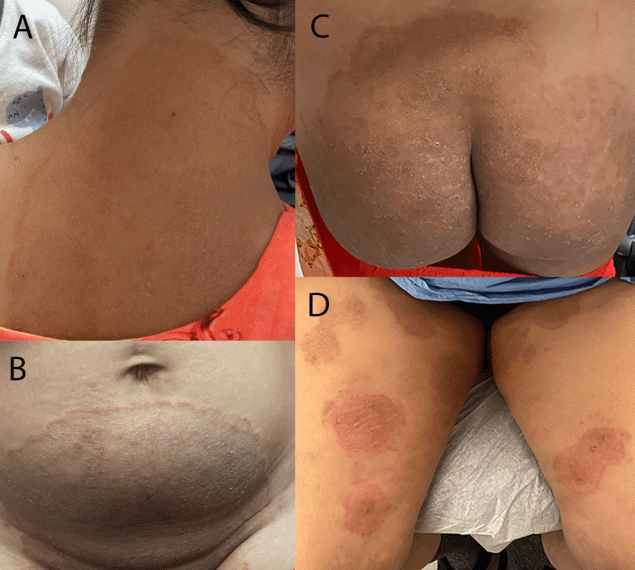
FIGURE. Lesions occurring on two patients with first reported U.S. cases of tinea caused by Trichophyton indotineae, on patient A’s neck, abdomen, and buttocks (A–C)* and on patient B’s thighs[SUP]†[/SUP] (D) — New York City, December 2021–March 2023
Photos/Lu Yin (A–C) and Vignesh Ramachandran (D). Used with patients’ permission.
* At initial dermatology evaluation, patient A had large, annular, scaly, and pruritic erythematous plaques of the neck, abdomen, groin, and buttocks.
[SUP]†[/SUP] At initial dermatology evaluation, patient B had widespread, discrete, scaly, annular, pruritic plaques affecting the thighs and buttocks.
https://www.cdc.gov/mmwr/volumes/72/wr/mm7219a4.htm#suggestedcitation
DOI: http://dx.doi.org/10.15585/mmwr.mm7219a4
Caplan AS, Chaturvedi S, Zhu Y, et al.
Tinea is a common, highly contagious, superficial infection of the skin, hair, or nails caused by dermatophyte molds.* During the past decade, an epidemic of severe, antifungal-resistant tinea has emerged in South Asia because of the rapid spread of Trichophyton indotineae,[SUP]†[/SUP] a novel dermatophyte species; the epidemic has likely been driven by misuse and overuse of topical antifungals and corticosteroids[SUP]§[/SUP] (1,2). T. indotineae infections are highly transmissible and characterized by widespread, inflamed, pruritic plaques on the body (tinea corporis), the crural fold, pubic region, and adjacent thigh (tinea cruris), or the face (tinea faciei) (1). T. indotineaeisolates are frequently resistant to terbinafine, a mainstay of tinea treatment (1,3). T. indotineaeinfections have been reported throughout Asia and in Europe and Canada but have not previously been described in the United States (3).
On February 28, 2023, a New York City dermatologist notified public health officials of two patients who had severe tinea that did not improve with oral terbinafine treatment, raising concern for potential T. indotineae infection; these patients shared no epidemiologic links. Skin culture isolates from each patient were previously identified by a clinical laboratory as Trichophyton mentagrophytes and were subsequently forwarded to the Wadsworth Center, New York State Department of Health, for further review and analysis. Sanger sequencing of the internal transcribed spacer region of the ribosomal gene, followed by phylogenetic analysis performed during March 2023, identified the isolates as T. indotineae (Supplementary Figure; https://stacks.cdc.gov/view/cdc/127678). Activity related to this investigation was reviewed by CDC and was conducted consistent with applicable federal law and CDC policy. ...
FIGURE. Lesions occurring on two patients with first reported U.S. cases of tinea caused by Trichophyton indotineae, on patient A’s neck, abdomen, and buttocks (A–C)* and on patient B’s thighs[SUP]†[/SUP] (D) — New York City, December 2021–March 2023
Photos/Lu Yin (A–C) and Vignesh Ramachandran (D). Used with patients’ permission.
* At initial dermatology evaluation, patient A had large, annular, scaly, and pruritic erythematous plaques of the neck, abdomen, groin, and buttocks.
[SUP]†[/SUP] At initial dermatology evaluation, patient B had widespread, discrete, scaly, annular, pruritic plaques affecting the thighs and buttocks.
https://www.cdc.gov/mmwr/volumes/72/wr/mm7219a4.htm#suggestedcitation