Announcement

Collapse
No announcement yet.

New study finds low levels of MLV GAG sequences in samples from CFS patients

Collapse
X
 
  • Filter
  • Time
  • Show
Clear All
new posts

  • New study finds low levels of MLV GAG sequences in samples from CFS patients

    http://okeefe-lab.blogspot.co.uk/201...v-gag.html?m=1

    http://www.newmicrobiologica.org/PUB...2012/3/341.pdf
    XMRV and polytropic MLV-related virus have been controversially associated with chronic fatigue syndrome (CFS).
    Subsequent reports failed to detect XMRV and MLV-related virus in CFS patients, and the previous results have been
    interpreted as a massive laboratory contamination by mouse DNA sequences. Among 12 sequential CFS patients, two
    were positive for XMRV/MLV sequences. In contrast, 40 selected control subjects were negative. CSF patients and
    controls were negative for mitochondrial mouse-specific DNA sequences. These findings do not confirm the high frequency
    of MLV-related viruses infection in CFS patients, but also contrast the widespread laboratory contamination
    previously suggested.
    Key words: Chronic fatigue syndrome, XMRV, MLV-related virus, Polytropic MLV, Ecotropic MLV.
    _____________________________________________

    Ask Congress to Investigate COVID Origins and Government Response to Pandemic.

    i love myself. the quietest. simplest. most powerful. revolution ever. ---- nayyirah waheed

    "...there’s an obvious contest that’s happening between different sectors of the colonial ruling class in this country. And they would, if they could, lump us into their beef, their struggle." ---- Omali Yeshitela, African People’s Socialist Party

    (My posts are not intended as advice or professional assessments of any kind.)
    Never forget Excalibur.

  • #2
    Re: New study finds low levels of MLV GAG sequences in samples from CFS patients

    CII
    @CII722
    The Center for Infection & Immunity is dedicated to pathogen surveillance & grasping how gene-environment-timing interactions contribute to health & disease.
    New York City ·


    #XMRV/#CFS/#ME study is complete. Was accepted for publication last week. Is due for release on Sept. 18.
    ------

    The study will be published online in mBio, which is an open access journal. #FreeForAll #XMRV #CFS #ME

    Comment


    • #3
      Re: New study finds low levels of MLV GAG sequences in samples from CFS patients


      Gammaretroviruses related to murine leukemia virus (MLV) have variously been reported to be present or absent in blood from chronic fatigue syndrome/myalgic encephalomyelitis (CFS/ME) patients and healthy controls. Using subjects from New York State, we have investigated by PCR methods whether MLV-related sequences can be identified in nucleic acids isolated from whole blood or from peripheral blood mononuclear cells (PBMCs) or following PBMC culture. We have also passaged the prostate cancer cell line LNCaP following incubation with plasma from patients and controls and assayed nucleic acids for viral sequences. We have used 15 sets of primers that can effectively amplify conserved regions of murine endogenous and exogenous retrovirus sequences. We demonstrate that our PCR assays for MLV-related gag sequences and for mouse DNA contamination are extremely sensitive. While we have identified MLV-like gag sequences following PCR on human DNA preparations, we are unable to conclude that these sequences originated in the blood samples.


      Novel MLV-GAG sequences detected in blood samples of ME/CFS patients

      From the Hanson lab, this paper published today in PLoS ONE describes a study carried out with meticulous attention to detail and proper use of controls while analyzing ME/CFS patient samples for murine-leukemia related viruses. Although novel MLV-like GAG sequences were isolated and sequenced from the patient samples (as opposed to controls collected from the same sites) Lee et al. concludes that it is not possible to determine the origin of these sequences, although the samples were not contaminated with mouse DNA at a detectable level. First, bravo! to the Hanson group for being able to get anything published that might go against the current dogma regarding MLV-like viruses in human samples published -- I guarantee you that there are people with similar data whom don't want to torture themselves by attempting to publish. The fact is, that it seems somehow MLV-like nucleic acids somehow end up in patient samples quite a bit, albeit at exceedingly low levels. Theoretically, even if there was an MLV-type infection in a human, it would be rapidly eliminated (unless of course we don't know everything about the immune system yet). I wonder if it is possible that such viruses could hide somewhere in the body, not replicating much - perhaps killing host cells occasionally resulting in a release of (potentially degraded) viral nucleic acid that is ultimately detected in the blood stream. I haven't analyzed these sequences yet compared to those we have detected in tissue samples from BPH patients, but seeing as our sequences were recently published on the USPTO website, someone else will probably try that. The link to the Hanson paper is here --


      Also from Dr O'Keefe.....

      it is interesting that everyone, us included, seems to have much more trouble amplifying the env and LTR regions - if gag amplification was merely due to mouse contamination, why don't these regions amplify as well? Keeping in mind that tests for mouse DNA are coming up negative...this might suggest that there is more divergence from known MLV-type sequences in these regions - we managed to sequence the variable region of env from one patient -- the sequence was most similar to env from an MLV virus known to cause disease in mice in the wild -- which is pretty interesting if you think about it...

      Comment


      • #4
        Re: New study finds low levels of MLV GAG sequences in samples from CFS patients

        http://cii.columbia.edu/blog.htm?LOfRcb&forumid=331851


        CII Press Conference
        September 18th, 2012
        10:30 AM (EDT)


        ?Multicenter Study on Chronic Fatigue Syndrome/Myalgic Encephalomyelitis?

        The causes of chronic fatigue syndrome/myalgic encephalomyelitis (CFS/ME) have long eluded scientists. In 2009/2010, two high-profile papers linked the syndrome to infection with a mouse retrovirus called XMRV (xenotropic murine leukemia virus (MLV)-related virus) and murine retrovirus-like sequences (designated pMLV: polytropic MLV). Given that affected patients often have symptoms consistent with a chronic infection, this viral connection was deemed plausible, and the findings were celebrated as a major achievement for a complex disease that afflicts nearly 1 million in the U.S.
        However, follow-up investigations by several laboratories were unable to detect XMRV or pMLV in CFS patients.
        To definitively resolve this issue, the National Institute of Allergy and Infectious Diseases commissioned a large-scale, multicenter study under the auspices of the Center for Infection and Immunity at Columbia University?s Mailman School of Public Health, in partnership with the Centers for Disease Control and Prevention, the Food and Drug Administration, the NIH?s National Cancer Institute and Warren G. Magnuson Clinical Center, and others (see full list below).
        Investigators from the multicenter study on CFS/ME and XMRV/pMLV will discuss their findings and future directions.


        A live video will be posted below.


        For attendance at the event, members of the media should RSVP by 9:00AM on Monday, Sept. 17th to Nsikan Akpan (212-342-9051 | nea2107@columbia.edu) or Stephanie Berger, Mailman School of Public Health (212-305-4372 | sb2247@columbia.edu).

        When: Tuesday, September 18, 2012
        Press Conference begins at 10:30 AM
        (EDT)

        Who: W. Ian Lipkin, MD (Moderator)
        Center for Infection and Immunity (CII), Mailman School of Public Health, Columbia University
        Principal Investigator and Director of Center for Infection and Immunity
        John Snow Professor of Epidemiology
        Professor of Neurology and Pathology

        Harvey Alter, MD
        National Institutes of Health, Department of Transfusion Medicine
        Distinguished NIH Investigator
        Chief, Clinical Studies
        Associate Director of Research

        Mady Hornig, MD
        Center for Infection and Immunity, Mailman School of Public Health, Columbia University
        Director of Translational Research at CII
        Associate Professor of Epidemiology

        Judy Mikovits, PhD
        Mikovits Consulting

        Francis Ruscetti, PhD
        National Cancer Institute, Frederick National Laboratory for Cancer Research
        Head, Leukocyte Biology Section
        Senior Investigator


        Collaborating Research Groups:

        Department of Transfusion Medicine, Warren G. Magnuson Clinical Center, National Institutes of Health, Bethesda, MD.
        Mikovits Consulting, Oxnard, CA.
        Division of HIV/AIDS Prevention, Centers for Disease Control and Prevention, Atlanta, GA.
        Cancer and Inflammation Program, Frederick National Laboratory for Cancer Research, Frederick, MD.
        Tissue Safety Laboratory, Office of Cellular, Tissue and Gene Therapies, Center for Biologics Evaluation and Research, Food and Drug Administration, Bethesda, MD.
        Nova Southeastern University, Fort Lauderdale FL.
        Miami Veterans Affairs Medical Center, Miami, FL.
        Brigham and Women?s Hospital, Boston, MA.
        Infectious Disease Clinic, Stanford University, Palo Alto, CA.
        Fatigue Consultation Clinic, Salt Lake City, UT.
        Levine Clinic, New York, NY.
        Simmaron Research Institute, Incline Village, NV.
        Department of Biostatistics, Columbia University, New York, NY.
        Department of Molecular Biology and Genetics, Cornell University, Ithaca, NY.
        Center for Infection and Immunity, Columbia University, New York, NY.
        Department of Molecular Biology and Microbiology, Tufts University, Boston, MA

        Comment


        • #5
          Re: New study finds low levels of MLV GAG sequences in samples from CFS patients

          The results of the multicenter study on XMRV/pMLVs in CFS/ME will be posted online in the open access journal mBIO at 12:01 a.m. Tuesday and the press embargo lifts at that time. Dr. Vincent Racaniello, Columbia virology professor and host of "This Week in Virology," will post a special episode of TWiV featuring Dr. Ian Lipkin, coordinator of the study, at 12:00 p.m. on Tuesday. TWiV site URL: http://www.twiv.tv/

          Comment


          • #6
            Re: New study finds low levels of MLV GAG sequences in samples from CFS patients

            Thank you, Summerflower. I'm expecting XMRV to be ruled out, but hope the effort might be helpful in other ways. Things got very heated regarding XMRV once it was discovered that the virus had evolved in a laboratory.
            http://www.ncbi.nlm.nih.gov/pubmed/22818188
            Curr Opin Virol. 2012 Aug;2(4):499-507. Epub 2012 Jul 19.
            Recombinant origin, contamination, and de-discovery of XMRV.
            Delviks-Frankenberry K, Cingöz O, Coffin JM, Pathak VK.
            Source

            Viral Mutation Section, NCI, HIV DRP, Frederick National Laboratory for Cancer Research, Frederick, MD, United States.
            [snip]
            XMRV is a laboratory-derived virus generated in the mid 1990s through recombination during passage of a prostate tumor xenograft in immuno-compromised mice. While these developments are disappointing to scientists and patients, they provide a valuable road map of potential pitfalls to the would-be microbe hunters.
            So while I think it has been valuable to be aware that potentially human infective retroviruses can evolve like this and contaminate laboratories, it looks like the virus was contained in laboratories.

            Also, the XMRV timing wouldn't be right as far as including the initial CFS/ME outbreaks in the 1980's. The Incline Village area is the same general habitat, I think, as where the hantavirus outbreak is occurring.


            So I still wonder about something zoonotic carried by mice being involved in CFS/ME.

            <object width="420" height="315"><param name="movie" value="http://www.youtube.com/v/AW0x9_Q8qbo?version=3&amp;hl=en_US"></param><param name="allowFullScreen" value="true"></param><param name="allowscriptaccess" value="always"></param><embed src="http://www.youtube.com/v/AW0x9_Q8qbo?version=3&amp;hl=en_US" type="application/x-shockwave-flash" width="420" height="315" allowscriptaccess="always" allowfullscreen="true"></embed></object>
            Last edited by Emily; September 17, 2012, 02:00 PM. Reason: Typo fix.
            _____________________________________________

            Ask Congress to Investigate COVID Origins and Government Response to Pandemic.

            i love myself. the quietest. simplest. most powerful. revolution ever. ---- nayyirah waheed

            "...there’s an obvious contest that’s happening between different sectors of the colonial ruling class in this country. And they would, if they could, lump us into their beef, their struggle." ---- Omali Yeshitela, African People’s Socialist Party

            (My posts are not intended as advice or professional assessments of any kind.)
            Never forget Excalibur.

            Comment


            • #7
              Re: New study finds low levels of MLV GAG sequences in samples from CFS patients

              Thanks Emily.

              Yes the researchers who said they were finding XMRV in the blood of CFS patients have said they were in fact not looking at 'XMRV'

              they say they realized that this experiment did not in fact show XMRV but proteins from a broader family of gammaretroviruses.
              The Western blot assay they used in Lombardi et al. just so happened to cast a wider net that uncovered proteins from any member of the gammaretrovirus family.


              ------------------------------


              May 30, 2011

              Dr. Bruce Alberts
              Editor-in-Chief
              Ms. Monica Bradford
              Executive Editor Science
              1200 New York Avenue, NW
              Washington, DC 20005

              Re: Lombardi et al.

              Dear Dr. Alberts and Ms. Bradford:

              As the corresponding author of the Lombardi et al. study I want to express my deepest concern about the proposed issuance of your editorial expression of concern regarding our XMRV findings and its association with chronic fatigue syndrome. This is especially so in light of the gross disregard for the integrity of the scientific process by the apparent willful breach of your embargo by one of the authors or their collaborators. This has resulted in the apparent public knowledge of the contents of your request that we retract our seminal paper. I would respectfully ask that you focus on the following key facts and reconsider your position. We share your deep concern over the number of negative non-replication studies in this new area of research. However, the publication of your editorial expression of concern over the validity of Lombardi et al. findings are premature and would have a disastrous impact on the future of this field of science. Please do not proceed down a path that could be detrimental to the scientific exploration of human retroviruses in infectious disease, cancer, and, therefore, the future health of millions around the world.

              First, the title and substance of the Lombardi et al. study Detection of an Infectious retrovirus, XMRV, in blood cells of patients with Chronic Fatigue Syndrome is accurate, and not one reported study has been able to show why it is not. Using four different methods including PCR (of cultured and co-cultured cells), detection of human gammaretroviral (HGRV) viral proteins (culture and co-culture detected by Western Blot and flow cytometry), anti-gammaretrovirus Env antibodies in human serum (competed by 7C10 rat monoclonal antibody), and virus isolation from primary cell and co-cultures, we reported evidence of human gammaretrovirus infection in at least 67 out of 101 CFS patients. In addition, we reported that 3.7&#37; of the control population had evidence of infection.

              Second, this significant study was conducted over eight months and conducted in five different laboratories. It resulted in the first isolation of a human gammaretrovirus from the blood of humans and concluded that this virus may be a contributing factor in the pathogenesis of CFS. Electron micrographs of gammaretroviruses isolated from patients cells were shown in the Lombardi et al. study and support these conclusions. These electron micrographs do not show VP62 plasmid contamination. In addition, we have maintained the viral isolates of five patients from which the electron micrographs were derived. Moreover, data presented in Lombardi et al. suggested additional strains of gammaretroviruses and viral Gag proteins could be directly immunoprecipitated from the blood of patients supporting a finding that additional strains of HGRV were isolated. In fact, subsequent work by Jones et al. presented at Cold Spring Harbors supports the presence of more than one strain of HGRV. The original manuscript submitted to Science discussed DG75, a human B cell line, from which a MLV-related virus was fully sequenced. This raises the possibility of many viruses originating from recombination events as human tissue has been passed through mice for more than five decades. PCR would not have detected a DG75 isolate but the rat monoclonal Env antibody used in these studies can detect DG75 isolates. This raises the question of how many gammaretroviruses are circulating in the human population with the potential of contributing to human disease.

              Third, this study showed that human gammaretrovirus was transmissible, and a more recent study has confirmed these data in an animal model and has shown that there could be different routes of entry and difference in blood reservoirs between acute and chronic infection.

              Fourth, this study conducted the following tests to insure that the reported data were not as a result of contamination, including the detection of a human antibody response to the virus, the screening of all reagents and cell lines for any evidence of gammaretrovirus contamination, human or otherwise. The antibodies used to detect viral proteins in Lombardi et al. were rat monoclonal and goat polyclonal antisera, all of which were negative for murine contamination; all reagents used in PCR and tissue culture were lot tested for contamination. In addition, it was clearly stated in the Supplemental Methods which taq enzyme manufactures were shown to be contamination free. None of the negative papers, which demonstrated contamination, used the enzymes or antibody reagents used and recommended by Lombardi et al.

              All samples and controls were processed in the exact same way and placed in a clean lab free from any other cell line. Only five human cell lines were grown in the WPI laboratories during the time these studies were conducted: Raji, SupT1, HFF, LNCaP and HSB2 and all were shown at the initiation of and throughout these studies to be free of XMRV/VP62 and all were used as negative control tested weekly by every method (including pelleting of supernatant over glycerol for virus isolation).

              No murine cell line was grown in the WPI labs prior to the submission of the Lombardi et al. manuscript. The murine BAF cell line was cultured and used after the July 22, 2009 NCI closed meeting on XMRV during which all of the data of Lombardi et al. was shown not only to one of the reviewers of the original manuscript but also to John Coffin who wrote the accompanying commentary. We were requested at that time to run the mouse mitochondrial assay to show absence of mouse contamination. We conducted this assay on samples from all 101 patients in Lombardi et al. and published these data in the subsequent Virulence addenda, a copy of which is attached hereto. While we have been advised by you that Paprotka et al. suggest a recombinant origin of an XMRV, it says nothing about the human gammaretroviruses detected and isolated from patient samples in Lombardi et al. They cannot have any data to support the conclusion “that laboratory contamination with XMRV produced by a cell line (22Rv1) derived from these early xenograft experiments is the most likely explanation for detection of the virus in patient samples.” In fact, the authors of this paper know full well that this explanation cannot explain XMRV integration in human tissue, in situ hybridization, or antibodies reported in prostate cancer or CFS patients. Furthermore, all strains of wild rodents have not been examined and other examples of ancestral XMRV can be found. Neither 22Rv1 nor any of the cell lines reported to be contaminated with XMRV or cell lines growing the VP62 infectious molecularly cloned virus was in the laboratories where the patient cells were isolated. This can also not in any way explain the Env antibodies demonstrated in patient plasma in Lombardi et al. The reactivity demonstrated to Spleen Focus Forming Virus (SFFV) Env was competed by the rat monoclonal antibody which detects all known xenotropic, polytropic and ecotropic MLVs. This again suggests that we have, in fact, detected more than one strain of human gamma retroviruses in these patient samples. Clearly data presented in Lombardi et al. where samples were PCR negative but Western blot positive, using the 7C10 antibody, further support the notion of a family of gammaretroviruses. These data must be appreciated as a complete body of evidence and not in the context of individual pieces, such as PCR amplification using primers designed to an arbitrary reference strain.

              All of these data led Harvey Alter, in the NIH State of the Knowledge Workshop (April 2011), to draw the conclusion that there existed no evidence of contamination in ‘either the Mikovits or Lo labs’.

              The authors are aware of ten negative CFS papers listed in PubMed on the subject of XMRV. Most of the negative studies failed to find any evidence of XMRV in any sample type. This would suggest that the methods and materials used in the non-replication studies are insufficient to use when attempting to detect human gammaretrovirus in the blood of human samples. The methods, processes, and materials of Lombardi et al. need to be followed precisely. The Alter and Lo study is the only study which has attempted a partial replication of the methods and materials of the Lombardi study, which confirmed evidence of MLV related viruses. Studies using multiple different methods are not replication studies, and studies optimized to detect murine gammaretroviruses and not human gammaretroviruses must be seriously questioned. See, Virulence attachment.

              Scientific research of human gammaretroviruses is in its infancy. Studies of XMRV and macaques are beginning to reveal information concerning the life cycle of the virus, multiple tissue reservoirs, and a description of factors that induce viral activation. These studies are critical to understanding gammaretroviruses in human disease. Other human studies such as the one by Fischer et al., reported the detection of XMRV in the respiratory tract of immunocompromised individuals pointing to the potential for gammaretroviruses to be more easily transmitted than all other known retroviruses. WPI researchers are contributing to the development of more accurate clinical testing methods with others in the blood working group. Without the participation of Drs. Alter, Lo and the WPI, who have proven gamma retroviral detection methods, it may be impossible to discover whether or not gammaretroviruses are a threat to human transfusion and transplantation medicine.

              In summary, human retroviruses are not known to infect individuals according to their sex or age therefore there can be no excuse as to why it would be acceptable to study the viruses in cancer but not in those with infectious neuro-immune diseases. They create lifelong infection in their hosts by integrating into the genome of their victims. Thirty years of murine gammaretroviral research provide compelling evidence that these viruses cause immune deficiencies, neurological disease and cancer in mammals and are therefore possible contributors to human neuro-immune diseases such as CFS. However, good scientific work is difficult and takes time. These ongoing studies deserve to receive a fair and impartial evaluation in the peer-review process. The critical question which remains is not simply whether gammaretroviruses play a role in CFS or cancer but in how many other human diseases? Therefore, we feel this is an extremely premature action which is not in the best interest of the scientific community or human health and again we respectfully request that you allow the scientific process to run its course unhindered by bias.

              Thank you for your thoughtful consideration of this matter.

              Sincerely yours,

              Dr. Judy A Mikovits
              Director of Research Whittemore Peterson Institute

              Comment


              • #8
                Re: New study finds low levels of MLV GAG sequences in samples from CFS patients

                The Lipkin XMRV / MLV CFS Study results are in.

                http://www.eurekalert.org/pub_releas...-vnt091312.php


                Chronic Fatigue Syndrome Is Not Linked to Suspect Viruses XMRV or pMLV.

                So does that mean all the findings by Dr O'Keefe (above) are incorrect and all the positive studies that found the viruses in prostate cancer were also a mistake?

                Comment


                • #9
                  Re: New study finds low levels of MLV GAG sequences in samples from CFS patients

                  Some more details from Lipkins Study......


                  None of the laboratories found evidence of XMRV or pMLV in samples from the recruited CFS/ME or control subjects. For quality assurance of the molecular tests, separate positive controls (blood samples intentionally spiked with XMRV/pMLV) and negative controls (blood samples prescreened and lacking the retroviruses) were used and confirmed that the diagnostic assays were functioning properly.


                  Nine control and nine CFS/ME blood samples were positive for XMRV/pMLV-reactive antibodies. The accuracy of this assay cannot be determined because there are no positive controls in the general population with XMRV serology. Nonetheless, there was no correlation of antibody reactivity in blood from CFS/ME and controls.

                  Comment


                  • #10
                    Re: New study finds low levels of MLV GAG sequences in samples from CFS patients

                    (LINK) http://news.sciencemag.org/scienceno...mr.html?ref=hp

                    Yesterday the paper which showed that XMRV was even discovered was retracted.


                    However the authors don't agree:

                    "But the discovery of XMRV, a new virus, still stands," says one of the authors, Robert Silverman of the Cleveland Clinic in Ohio, and an erratum to withdraw the prostate cancer link would have sufficed. "Why retract results that are valid?" Silverman asks.

                    In the paper, the group writes that XMRV is still a "genuine infectious agent" with "as-yet undefined pathogenic potential"; they point out that the virus is able to infect mice and two primate species and that several research groups have discovered interesting biological properties that could be useful, for instance, in cancer research.

                    Comment


                    • #11
                      Re: New study finds low levels of MLV GAG sequences in samples from CFS patients

                      Originally posted by Summerflower View Post
                      (LINK) http://news.sciencemag.org/scienceno...mr.html?ref=hp

                      Yesterday the paper which showed that XMRV was even discovered was retracted.


                      However the authors don't agree:
                      That's a very interesting article, Summerflower. So historically it is unprecedented to totally retract papers reporting the discovery of viruses. Even the mechanism of the retraction sounds odd:

                      "This is an unusual case," confirms Arturo Casadevall, a microbiologist at Albert Einstein College of Medicine in New York City and the editor-in-chief of mBIO. "Usually the authors are involved in the decision and on most retractions at least some of the authors agree with the retraction." Guidelines from the Committee on Publication Ethics stipulate that for retractions, "[w]henever possible, editors should negotiate with authors and attempt to agree a form of wording that is clear and informative to readers and acceptable to all parties."
                      I wonder if the XMRV-contaminated cell cultures will continue to be used in research?
                      _____________________________________________

                      Ask Congress to Investigate COVID Origins and Government Response to Pandemic.

                      i love myself. the quietest. simplest. most powerful. revolution ever. ---- nayyirah waheed

                      "...there’s an obvious contest that’s happening between different sectors of the colonial ruling class in this country. And they would, if they could, lump us into their beef, their struggle." ---- Omali Yeshitela, African People’s Socialist Party

                      (My posts are not intended as advice or professional assessments of any kind.)
                      Never forget Excalibur.

                      Comment


                      • #12
                        Re: New study finds low levels of MLV GAG sequences in samples from CFS patients

                        Hi Emily, thought this might interest you, I found there was actually a XMRV / MLV study that took place in the United Kingdom. This UK Study was carried out by Francis Ruscetti and his team at the NCI. The Blood samples were taken from 50 CFS patients who lived in the London area.

                        ---------------------------


                        210
                        Agenda Item: U.K. study

                        We'll move to the third talk in this section. It's a U.K. study. Dr. Mikovits will present this.
                        DR. MIKOVITS: Thank you. Today I have been asked to talk about a study we presented at the XMRV workshop for a group of patients in the U.K. Since the first isolation of XMRV from the blood of CFS patients, my collaborators and I at the NCI, SAIC, and Whittemore Peterson Institute have been working to develop more sensitive assays for detection of infectious virus. The rationale for these studies is that in our work over the past year we have developed more sensitive methods for both the biological and molecular amplification of human MLV-related viruses, which in this talk will be called HMRV, in the blood and plasma. We have developed these technologies. These methods were used to determine the incidence of HMRVs in a U.K. cohort of ME/CFS. Importantly, this cohort was diagnosed using the more rigorous Canadian Consensus Criteria. Those are the criteria that Tony Komaroff and the study you heard from Dr. Lo, David Bell, as well as Dan Peterson from the original Science paper, used throughout their patient populations. It's important, because of the heterogeneity of the disease that we heard from Dr. Hewlett earlier, that the cases were similar.

                        Let's talk a little bit more about the study cohort we used from the London area of the U.K. They had gotten diagnosis of what they call myalgic encephalomyelitis. Or, often, post-viral fatigue is how it's diagnosed in the U.K. primarily. All of these patients do meet the Canadian Consensus Criteria of CFS that are used in those studies that we talked about today and in our original study and throughout the WPI. The disease duration in this patient population was 9 to 26 years, with greater than 50 percent of the patients actually housebound, and many bedbound. The onset of disease could often occur in childhood or puberty. We won't go into the possible reasons for that today. In addition to that profound post-malaise/fatigue that is really the sine qua non diagnosis of CFS that meets the Canadian Consensus Criteria, the other symptoms included severe cognitive dysfunction, multi-joint pain, the onsets of new and frequent migraine headaches, vertigo, dizziness, lymphadenopathy, profound mitochondrial dysfunction, which might explain the energy. Many of these patients have GI disturbance and dysbiosis, an inability to absorb nutrients, and medications as well. They have chronic infections. As we have often heard, well, those CFS patients have everything. Yes, they do, and that's the point. A healthy immune system doesn't have chronic EBV or chronic HHV-6. We see shingles in a 30-year-old often in these patient populations.

                        Importantly, many report a flu-like onset. They knew the day they got sick, and didn't recover. The current age of the study participants that were used was 19 to 70. Interestingly, in the 50 that were done randomly in this first pilot study, equal numbers were male and female. The study design we used is similar to that which we used in other studies done at the WPI. We have the blood drawn by Phlebotomy Services International, which is an independent certified phlebotomist group that goes around the world. PSI codes and ships those samples. In this case they were shipped to the NCI, where they were processed in a laboratory that had no previous XMRV work nor any previous murine research -- a human lab that had not done XMRV research previously. The plasma and the PBMCs were isolated two days after the blood collection. That was largely a matter of the shipping of the samples from the U.K. All the samples were tested in two independent labs, blinded. We blinded in 50 healthy controls taken from blood donors by our collaborator, Jonathan Kerr in London, in the mid-2000s, 2005 to 2008. We didn't have fresh draws from those blood donors, but these were blinded into the study, as those were the controls that we had available.

                        The samples were tested for the four methods. I'll go through it very carefully:

                        For plasma XMRV RNA.
                        For cell-free transmission from the plasma to the LNCaP cells, which we have heard about earlier. I'll describe that assay in detail.
                        We looked for plasma antibodies to HMRV viral proteins.
                        We Western-confirmed the positive cases from those transmission studies. Finally, we did sequence characterization of the HMRV isolates.

                        First, we'll talk about the plasma PCR. We had never before done direct plasma PCR. We had been working with the Blood Working Group and thought that perhaps delayed processing, which had been done in other studies, might increase our ability to see viral RNA which may have been associated with other blood components and actually released into the plasma. When we did this plasma RNA from 140 &#181;L of the plasma from 48 percent or 24 percent of the patients, we could see in this top, using the Lombardi nested primers and conditions, a very strong band for the gag. These were all sequence-confirmed to be gag of XMRV, but this amplicon wouldn't distinguish the polytropic sequences. It was very small.

                        Secondly, we used the Lo primers, went back to these samples that had not been previously frozen and thawed, because we aliquot them into .5 mL aliquots. You can see that two patients who were negative using our PCR were positive using the PCR protocol of Dr. Lo, suggesting that that also sequenced. But we didn't see it as polytropic. Maybe that's just our phylogenetic analysis. But all were confirmed by sequences and again highlight that subtle differences in PCR protocols can give you really big differences in results, as you would have found far fewer of these patients positive by the Lo protocol. Importantly -- and, unfortunately, it doesn't show well here -- we used 5 femtograms of DNA from the murine cell line that we have in the lab. We could see no mitochondria-specific amplicon, as Dr. Lo described.

                        The other thing that we do in all of our studies you see here number 2767 -- those are patients from the original WPI Science study, where we consistently and over time -- over three years' time -- can both detect plasma viremia and isolate from that patient. We carry this sample throughout these studies. We do that with several samples in every study. The control samples, as I mentioned, that we blinded in from Dr. Kerr -- you can see that very few actually had XMRV or HMRV RNA in the plasma. But importantly, two out of 50 that were reportedly from the healthy blood donors -- was 4 percent of the population there in the U.K. I do want to remark that of the two negative studies that had come out at this time from the U.K., they had absolutely zero incidence in controls or patient population of XMRV.

                        Since we are not a PCR lab and neither we nor the Science paper nor our work focuses on PCR, we went to doing the culture techniques that you have heard about today. I'll describe them in a bit more detail to show the isolation and characterization -- that these were indeed representing infectious virus. In the assay that we used in the Lombardi study, shown in the top line here, we take plasma or activated -- this is dividing peripheral blood and mononuclear cells -- from the patients, and we co-culture them on the prostate cancer cell line, which was responsive to androgens and inflammatory cytokines. This is important because we know we have characterized the LTR, in Steve Goff's lab and Bob Silverman's lab, and we know that there are hormone-responsive elements there that would be an on switch to make the virus replicate more in the cells that were responsive to androgens.

                        We culture these for 21 to 42 days. Dr. Hanson mentioned four passages. This is a lot of cultures. Carefully looking at other negative studies, they might culture them for a week. We follow them, in addition, to PCR by Western blots from antibodies. These are monoclonal antibodies which were described in the original Science paper. This rat monoclonal to the envelope of this spleen focus-forming virus, which is a polytropic, xenotropic virus -- importantly, this antibody was characterized by Sandy Ruscetti all the way back in 1982. But this surface unit I show you here in the Western for the transmission of three of these U.K. patients -- this antibody recognizes all polytropic, xenotropic, and ecotropic viruses. This antibody -- and that may be why our numbers were so high, because our original paper didn't originally rely on just the PCR, when, in fact, this antibody could detect all of the viruses. I'll refer you to that paper. If you look at Figure 1, you will see PCR-negative patients who clearly we could culture virus in, detect it from the antibody, and sequence whole virus.

                        This is the assay, which is quite labor-intensive and cumbersome. You know that it took us quite a while to do these 50-odd samples and 50 controls. We have been developing -- and you heard this from Dr. Le Grice at the last BPAC meeting in July, so I won't go into detail -- an assay in which an MLV vector has an inactivated green fluorescent protein in it. That vector is packaged by either XMRV or any MLV-related virus. You then infect those cells with that virus, if it's in the plasma, and in only 4 to 18 days, you can see green cells, representing infectious virus. In order for this vector to go from inactive to activated, it needs both reverse transcriptase and integrase. So it's important that this assay is an assay for infectious virus.

                        We show here that you can also quantitate it by flow cytometry and clearly see and count the green cells. Hopefully, that has been speeding things up a lot. When we use it in the U.K. samples, here is a positive control. Only 11 percent are positive. But that is due to viral interference and other things about this assay. But clearly a negative and clearly a positive. Both of these samples, if you go back and look at that first figure, were plasma PCR-positive, suggesting, but not proving, that it's infectious virus. Now we can see that 78 percent, 39 out of the 50, were positive in this infectious assay. This is just showing you other numbers that were negative and positive in the same assay, as you can see here. When we confirmed all of the samples -- and I show you here only the positive -- we confirmed by Western analysis, using an anti-MVL envelope. This is a xeno, so it is not the monoclonal I showed you, and then a Gag antibody as well. You can see that we can detect both Gag proteins and envelope proteins in these Western analyses, confirming that we had, in fact, transmitted the virus from the plasma of these patients to the LNCaP -- we call these DERSE cells.

                        Importantly, I show you that 2767 positive control that we carry throughout these studies. We next amplified a wider range, shown in the box on the top, of the envelope. When you look at small amounts of envelope, maybe due to the diversity that is wider than we originally anticipated -- when we actually did a PCR in the pol-pro region, extending down 600 base pairs of product into envelope, and then we sequenced -- I show you here, representative, three of these U.K. samples we could see that they were indeed more similar to XMRV than to the polytropic viruses we have been hearing about this morning.

                        Interestingly, this patient, U.K. 1023, was negative in all of the other assays, but we could actually, from the LNCaP, which was a DERSE assay -- it was only 2 percent, but we could actually see by Western that there was indeed virus there, and we could clone it out. We are doing full-length sequencing of as many of these viruses as we can from single cells at this time.

                        We next talk about the serology assay in these patients. This is the assay that you heard from Maureen Hanson and that was described in detail in the Science paper. We use a cell line that expresses the murine spleen focus-forming virus envelope, that same region that I showed you. The antibody recognized all known polytropic, xenotropic, and ecotropic viruses. We take a plasma from a patient sample and incubate it with the non-expressing cell line. You see nothing in this histogram, which shows increasing fluorescence and density of the binding of a secondary fluorescently labeled IgG. But here, with the patient sample on the envelope-expressing cell line -- clearly suggested that there is antibody to the envelope in the patient's plasma. You can compete that using that monoclonal antibody. If we co-culture the monoclonal antibody with the patient sample, you see that you can compete either at 1-to-10 or 1-to-50, demonstrating the specificity of this antibody and that indeed the patient samples do contain antibodies to spleen focus-forming virus envelope.

                        This is how the shift looks. We did this in all of the patient samples and controls. When we compare the detection of antibody reactivity with virus isolation from the plasma, you can see a concordance there in essentially all of them. There were only five samples where we could isolate virus, but could not detect presence of an antibody -- we don't know why that is -- and a few samples where we could detect antibody and not actually isolate virus. In summary, then:

                        We could detect gag in the RNA in the plasma in 58 percent of the 50 patients.
                        We could transmit 78 percent of the patient samples to the LNCaP cells.
                        We see antibody reactivity in 68 percent of those 50 patients.
                        We could sequence the envelope products, showing that the predominant HMRV in this U.K. cohort is indeed XMRV.

                        We conclude that multiple methods are necessary to detect evidence of XMRV infection. In this case, in a very well-defined cohort, similar to the positive studies, we could detect it in greater than 70 percent. With that, I'll thank my collaborators and funding and you for your attention. DR. HOLLINGER: Thank you.

                        Questions?
                        DR. COFFIN: The virus is growing out in the DERSE cells. How much of that have you sequenced?
                        DR. MIKOVITS: We have sequenced large parts of the envelope and the gag. We --
                        DR. COFFIN: I'm just curious, because one expectation in those cells is that what might happen -- the virus that grows out may not actually be the virus that you originally started with.
                        DR. MIKOVITS: That's why we -- DR. COFFIN: It could be a recombinant that has picked up useful sequences from the vector, like the LTR. It would be nice to see if that's happening.
                        DR. MIKOVITS: Indeed, and that is why we also run those LNCaPs without that vector. We have been sequencing the virus out of the LNCaPs where I showed you the Westerns. We run both assays, because we recognize that that might happen. It would be interesting if it did indeed happen.
                        DR. RUSCETTI: Can I follow up on the Coffin question?
                        DR. HOLLINGER: Could you give your name?
                        DR. RUSCETTI: Ruscetti, NCI.
                        We have done LNCaP and the DERSE cell on several isolates and found no difference, at least in the envelope and gag regions.
                        DR. COFFIN: I was thinking particularly about the LTR, which might well have been exchanged.

                        DR. RUSCETTI: We are just beginning to work to look at the LTR. We have been pushed into it by Jonathan, who asked us at every meeting to look at it. So we are now looking at it. But we don't have any results for it.
                        DR. MIKOVITS: And it could well be a key to the reservoir. The LTR is really a key, maybe, why we can't grow this virus in these cells. We are looking at other cell types right now.
                        DR. COFFIN: That harks back, in a sense, to the other talks. To my knowledge, nobody has ever grown a virus that has a polytropic or a modified polytropic-type LTR in it, unless somebody in the room has done that recently and I haven't heard about it. DR. MIKOVITS: Usually these are seen with xenotropic, actually mobilizing the polytropic. The polytropic indeed becomes the pathogenesis. But they need the xenotropic to be --
                        DR. COFFIN: In mice that seems to be the case.

                        DR. NELSON: These are pretty convincing data, to me, that this infection is real. You showed antibodies. One issue that remains is, is this the cause or is this the result? It's quite possible that they have something else - the immune system crashes or whatever -- and they get infected with whatever is around, a mouse or whatever. Have these patients been studied for other chronic viral infections, HHV-6, HTLV-2, or EBV? Have these patients had an extensive virologic/infectious disease workup?

                        DR. MIKOVITS: These patients in the U.K. have not. It is a psychosomatic disease in the U.K., and they can't get those types of medical treatments easily and maintain their benefits. In our study in Science, the answer is yes. These patients have multiple chronic active infections -- EBV, HHV-6, CMV, as I mentioned, shingles. We see everything -- mycoplasma. It looks to us like an AIDS patient, with an obvious hypothesis being that the retrovirus causes the underlying immune deficiency. But it alone can't cause the disease. It needs the co-pathogens. You can have HIV without having AIDS, but you can't AIDS without having HIV and one of 25-odd co-pathogens.
                        DR. NELSON: So it looks like the next step is to there are several repositories, and this disease may be frequent enough that one could identify an infection with this agent. You have 4 percent of blood donors or whatever. These people need to be followed. These people need to be followed to answer the question: Does this infection occur before the chronic fatigue syndrome or afterwards? If it occurs afterwards, then it's just a passenger; it isn't the case. I think that's a critical question.

                        There are some repositories, the NHANES, Washington County, Maryland, where there are large numbers of samples that are stored and frozen, to go back and look at incident disease after that. It seems like this should be of some priority at this point.
                        DR. MIKOVITS: It is a critical question, but we can't do it with the existing assays. Part of the reason we were developing these, hopefully, more high-throughput assays is so that we could do the large-scale epidemiological studies necessary. I agree completely.

                        Comment


                        • #13
                          Re: New study finds low levels of MLV GAG sequences in samples from CFS patients

                          Originally posted by Emily View Post
                          So I still wonder about something zoonotic carried by mice being involved in CFS/ME.
                          Yes I'm still wondering about that.

                          I noticed that in the Lipkin XMRV Study, 6&#37; of the study group (patients and controls) cross-reacted with a test designed to detect SFFV (Spleen Focus Forming Virus).

                          Comment


                          • #14
                            Re: New study finds low levels of MLV GAG sequences in samples from CFS patients

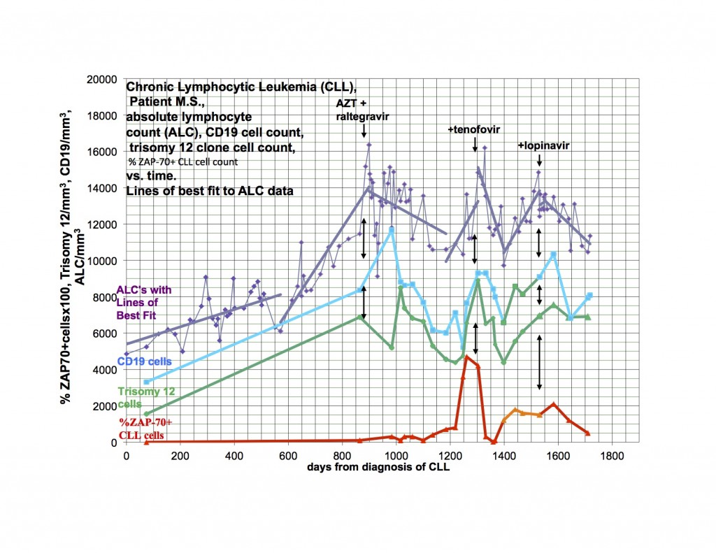
                            I also found this story about a doctor who has cancer. He tested positive in an XMRV / MLV research study and is now treating his Leukemia with anti-retroviral drugs.


                            I have CFS/ME and cancer. My data supports the presence and importance of retroviruses in the pathogenesis of both disorders and the potential for anti-retroviral drugs to help us.

                            My data shows the presence of a clonal T-lymphocyte expansion which is probably CD8 cells. Having a clonal T-lymphocyte expansion is abnormal. In addition, I have increased monocytes (monocytosis). I have looked at 56 of my patients with various types of cancer and half of them also have a detectable T-lymphocyte expansion and monocytosis. My point is that what I see in myself may be a common phenomenon and actually may be happening in millions of people. I haven’t looked in CFS/ME, because I limit my practice to Hematology-Oncology.

                            T-lymphocytes and monocytes are important because they are permissive of retroviral invasion and because they are pre-programmed to make cytokines. Presumably the increased numbers of these cells after infection has occurred would lead to increased amounts of cytokines that would dysregulate the immune system. In addition, monocytes give rise to the microglial cells which migrate to the brain and spinal cord and potentially could deposit abnormal amounts of cytokines in direct proximity to the neural elements.

                            Dr. Lipkin mentioned a polyclonal expansion of B-lymphocytes as important in our type of disorder and this could be a result of the increased amount of cytokines. In fact, in my patients who had a clonal T-lymphocyte expansion and/or monocytosis, 50&#37; had autoimmune markers. What is reasonable to postulate is that we now have explained the known connection between inflammation and cancer and neurodegenerative disorders and it all goes back to the retroviruses.

                            I have previously shown response of my leukemia, monocytosis and clonal T-cells to AZT, raltegravir and after relapse a second response to the addition of tenofovir. I had hoped to demonstrate that a PI would be valuable treatment and waited to add this category of drug until relapse. My leukemia responded and relapsed in parallel with the monocytosis and clonal T-cell expansion but at a somewhat different rate.

                            The last graph shows that the clonal T-lymphocytes, CD8 lymphocytes and monocytes were increasing after relapse on AZT, raltegravir and tenofovir. Quest discontinued doing the quantitative TCRγ in 2011. As a favor to me, Quest put it up but with different reagents so that the results from the new assay are not superimposable over 2011’s. Please note that the clonal TCRγ values are a ratio rather than an absolute number of cells.

                            I didn’t have the new clonal T-cell assay available until later in the graph and didn’t think of looking for CD8 cells initially. After the relapse was clearly documented, I added the PI, lopinavir. The T-cell parameters and monocytes continued to trend up for the next 4-8 weeks before clearly trending down.

                            The only reasonable explanation for what has happened to me and what I have found in half of my cancer patients is that retroviruses participate in the pathogenesis of our illnesses. Please note that I do not claim that the retroviruses cause the illnesses but emphasize that the retroviruses participate in the pathogenesis. The likelihood is that genetic susceptibility and toxic exposure determines whether an infected person would develop disease and how it would be manifested.

                            It is clear to me from the data I have, that many people have susceptibility to develop disease after infection with retroviruses. It is now up to the scientists to identify the infection and the basis of genetic susceptibility. Once there are easy methods to identify who is infected and with what, treatment trials could be started. We must go forward.

                            Michael Snyderman, MD

                            Comment


                            • #15
                              Re: New study finds low levels of MLV GAG sequences in samples from CFS patients


                              Plasmacytoid Dendritic Cells in the Duodenum
                              of Individuals Diagnosed with Myalgic Encephalomyelitis
                              Are Uniquely Immunoreactive to Antibodies
                              to Human Endogenous Retroviral Proteins


                              Comment

                              Working...
                              X