 Tweet
Tweet
Emerg Infect Dis
. 2020 Jun 22;26(9).
doi: 10.3201/eid2609.201806. Online ahead of print.
Persistence of Severe Acute Respiratory Syndrome Coronavirus 2 in Aerosol Suspensions
Alyssa C Fears, William B Klimstra, Paul Duprex, Amy Hartman, Scott C Weaver, Kenneth S Plante, Divya Mirchandani, Jessica Ann Plante, Patricia V Aguilar, Diana Fern?ndez, Aysegul Nalca, Aysegul Totura, David Dyer, Brian Kearney, Matthew Lackemeyer, J Kyle Bohannon, Reed Johnson, Robert F Garry, Doug S Reed, Chad J Roy
- PMID: 32568661
- DOI: 10.3201/eid2609.201806
Abstract
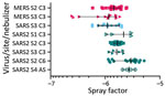
We aerosolized severe acute respiratory syndrome coronavirus 2 and determined that its dynamic aerosol efficiency surpassed those of severe acute respiratory syndrome coronavirus and Middle East respiratory syndrome. Although we performed experiment only once across several laboratories, our findings suggest retained infectivity and virion integrity for up to 16 hours in respirable-sized aerosols.
Keywords: 2019 novel coronavirus disease; COVID-19; MERS-CoV; Middle East respiratory syndrome coronavirus; SARS-CoV; SARS-CoV-2; aerosol; coronavirus diseases; respiratory diseases; severe acute respiratory syndrome coronavirus; severe acute respiratory syndrome coronavirus 2; viruses; zoonoses.
Comment